lung cancer in cats x ray
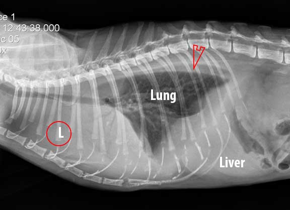
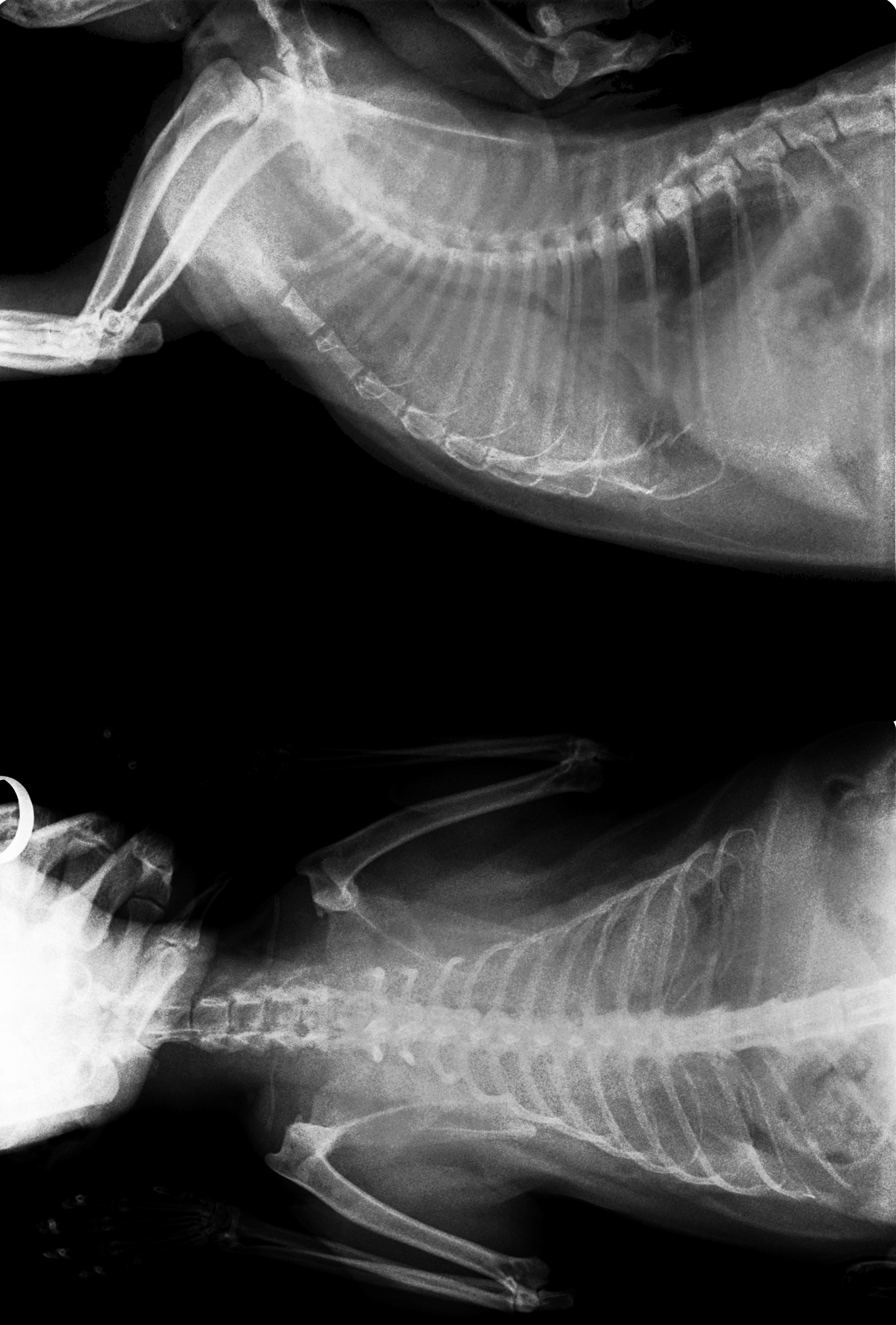
The top red arrow points to the aorta. Not All Dark Masses are Tumors.
Radiology Case Of The Week Feline Miliary Pulmonary Pattern
Lung tumors are considered rare in cats and dogs.

. Feline lung cancer is a cancer that is rare in cats. It is much more common for cats to develop a tumor in the lungs after cancer has spread from another location than for the tumor to start in the lungs. Pale or bluish lips gums and nose tissue.
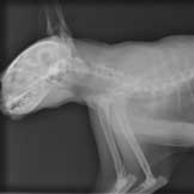
Ad Life Gold - Trusted Care For Pet Cancer. Symptoms of Cat Lung Tumors. This is a labeled normal feline DV dorsoventral chest X-Ray of a fat cat.
To evaluate respiratory conditions like asthma bronchitis and pneumonia heart conditions broken ribs and to look for fluid and tumors within the chest cavity. Lymphoma in cats is the most common. What Is Lung Cancer in Cats.
Chest radiographs x-rays are one of the screening tests done in both dogs and cats diagnosed with cancer to check for the presence of metastasis. A type of x-ray where the cat ingests barium sulfate followed by an x-ray. The first symptom of cat lung tumors is usually a mild cough similar to that associated with feline asthma.
Lung tumors in cats create symptoms similar to those of other. Your veterinarian may recommend an FNA and cytology or a biopsy to confirm the diagnosis and determine exactly what kind of lung cancer is present. This will ease the respiratory symptoms.
There are two types of lung cancer that affect cats. If your cat develops a tumor that is prone to metastasize to the lungs your veterinarian will likely recommend chest x-rays. Signs of an acute respiratory illness include labored breathing periodic blackouts or fainting.
Imaging methods to examine the lungs include chest X-ray low-radiation-dose chest Computed Tomography and standard-radiation-dose chest CT. If there is pleural effusion occurring in your cat the vet will then draw out the fluids with a needle. Most lung tumors are single masses that can be easily seen on routine X-rays of the chest.
X-rays of the chest are probably the single most useful diagnostic tool in making a preliminary diagnosis of lung cancer. Joanne Lynn Intile DVM MS DACVIM. All-Natural Support For The Best Quality Of Life.
7 This can be disastrous since it only takes an average of 136 days for a lung tumor to double in size. The signs of a chronic lung disease are. Not all pets with pulmonary tumors exhibit clinical signs and are often diagnosed incidentally from routine chest X-rays.
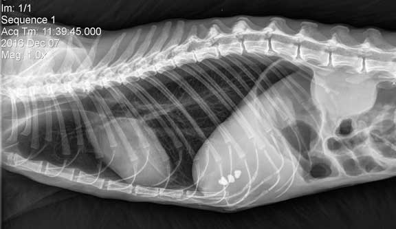
A thoracic chest radiograph X-ray is a procedure that allows your veterinarian to visualize tissues organs and bones that lie beneath the skin of the chest cavity in Cats. What causes lung cancer in cats. They had been referred to me for evaluation of a tumor in her chest.
For example if your cat. Lung tumors are considered rare in cats and dogs. Diagnosis of lung cancer usually starts with a chest X-ray.
X-rays also evaluate for the presence of fluid in the chest cavity the size of the heart and associated blood. Signs of lung cancer are hard to detect until it has reached a late stage while pneumonia symptoms. Thoracic radiographs are recommended for any cat or other pet with difficultly breathing or with suspicion of heart disease or lung disease.
Cats with certain health problems may get x-rays more frequently. Trixies owners sat stone-faced across from me in the exam room. The most common areas of the body that are x-rayed in cats are listed here.
Your veterinarian may be looking for signs of heart disease lung infections asthma tumors or metastatic spread of cancer. Stage 1 stage 2 and stage 3a lung cancers are considered treatable. They were a middle-aged couple filled with worry for their beloved 14-year-old tabby cat.
Many cats go their whole lives without ever needing an x-ray. A severe acute attack is likely to result in a cats death if veterinary treatment is not immediately available. Supports Your Pets Daily Comfort and Helps Maintain Their Energy Long-Term Vitality.
Pulmonary carcinomas have a high tendency to. To look for problems with the organs and space in the abdomen including. Small Cell Lung Cancer Chest X Ray - 17 images - diagnosing lung cancer non small cell lung cancer nsclc imaging practice essentials small cell lung cancer x ray stock image c013 1080 science photo imaging in small cell lung cancer overview radiography computed.
Certain breeds are more predisposed to develop pulmonary tumors than others. Because initial symptoms are mild misdiagnosis of cat lung cancer in its early stages is common. Ad Chewys 2-Step Process Makes it Easy To Fill Order Your Pets Prescriptions.
The fluid can then be. The barium coats the inside walls of the gastrointestinal which appear. Ultrasound-guided fine needle aspiration or biopsy will confirm the diagnosis.
The major goal of cancer treatment is to prevent metastasis. Ultrasound-guided fine needle aspiration or biopsy will confirm the diagnosis. Not all pets with pulmonary tumors exhibit clinical signs and are often diagnosed incidentally from routine chest X-rays.
Lung cancer on the other hand occurs when cells grow out of control and form tumors. To start pneumonia is a chest infection that causes inflammation to the air sacs in the lungs. Pneumonia pneumothorax pulmonary edema lung cancer primary or secondary lung abscess heart disease asthma pleural effusion pulmonary thromboembolism.
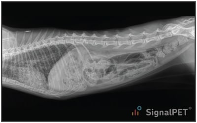
This is a normal cat lateral abdominal X-Ray. CT lung cancer screening has revolutionized medical imaging by providing more detailed information than conventional X-rays and ultimately offering better care for patients. The trachea and esophagus are also evaluated with this series.
Lung tumors may appear on chest X-rays as ill-defined lesions in the lung tissue. A 2019 review of 21 studies found that 20 to 23 of chest X-rays in people with lung cancer symptoms were falsely negative for lung cancer. The bottom red arrow points to the posterior vena cava bringing venous blood from the back of the body to the heart.
Chest X Ray To Diagnose Lung Cancer - 17 images - stages of lung cancer stages symptoms and diagnosis healthy chest x ray stock image p590 0229 science photo library small cell lung cancer treatment pdq patient version national pin on unusual x rays. However of the tumors that start in the lungs primary tumors nearly 80 of them are cancerous. In the later stages this cancer often spreads to the.
However x-rays cannot detect small tumors less than 3 millimeters in size so computed tomography CT scan may also be necessary. 35 Off Your First Autoship Easy Refills. Certain breeds are more predisposed to develop pulmonary tumors than others.
Most primary lung tumors are a type of cancer called. An x-ray of the chest will be called for and in the case of lung cancer the tumor will show up on the x-ray. However there are fundamental differences between the two.

Pin Page

Using Thoracic Radiographs To Differentiate Pulmonary And Cardiac Diseases In Dogs And Cats Medvet
Feline Radiographs X Rays
Learn How To Read A Cat X Ray Long Beach Animal Hospital

Pin On Dog Stuff

Image Gallery Primary Metastatic Tumors Part 1 Clinician S Brief Metastatic Tumor Thoracic Cavity

Lung Cat No 1 Diffuse Severe Bronchointerstitial Pattern Download Scientific Diagram

Dark Lung Spots On X Ray

Abnormal Shadow On Chest Radiograph Medpage Today

On Call Radiology Common Radiology Findings On Call And In The Emergency Room And During The Nightshift

Using Thoracic Radiographs To Differentiate Pulmonary And Cardiac Diseases In Dogs And Cats Medvet

Veterinary Radiography
Learn How To Read A Cat X Ray Long Beach Animal Hospital
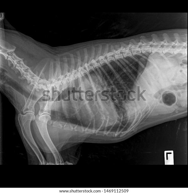
X Ray Lung Cancer Dog Stock Photo 1469112509 Shutterstock
File X Ray Of Felv Positive Cat With Lung Cancer Jpg Wikipedia

Feline Radiographs X Rays

Pin Page
Cancer In Cats Not All Dark Masses Are Cancerous Tumors Cancer In Pets Petmd

Learn How To Read A Cat X Ray Long Beach Animal Hospital